Leading Technology
Anatomage Tables-Virtual Lab
RESEARCH & TECHNOLOGY
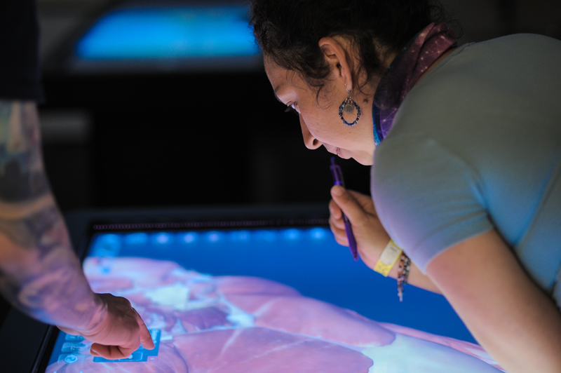
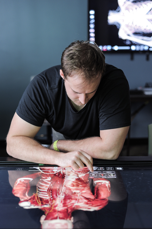
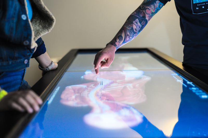
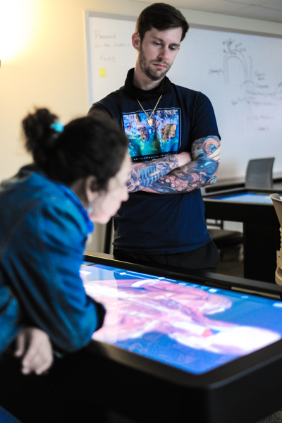
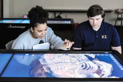
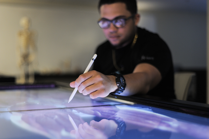
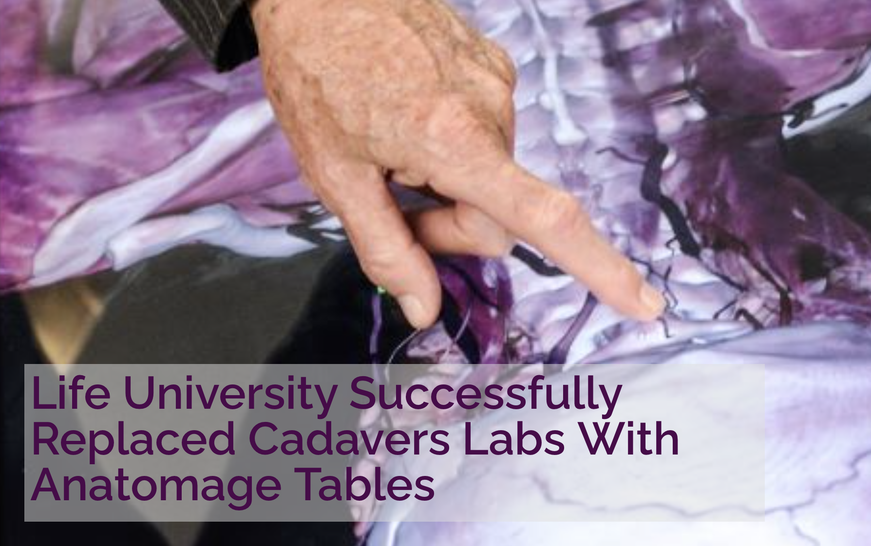
Life University’s Virtual Anatomy Lab was created in 2015 and allows students to study anatomy on state-of-the-art digital Anatomage tables, rather than cadavers. The Life University Virtual Anatomy Lab features more Anatomage tables than any other higher education institution in the world. Additionally, unlike cadaver dissection, the University’s Anatomage tables provide students with life-sized, multi-layered 3D technology as well as the ability to view CT scans, giving them a detailed understanding of a body’s systems and how they are interconnected.
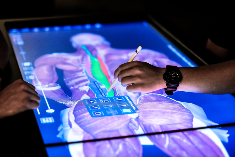
All of the Anatomage Tables at Life University have thousands of high-resolution 3D images taken from actual female and male human cadavers which allows students to label, highlight and capture detailed images to study at another time on their computer or tablet. And several research studies have shown that studying anatomy and physiology on Anatomage tables leads students to be more engaged and perform better academically compared to cadaver dissection.
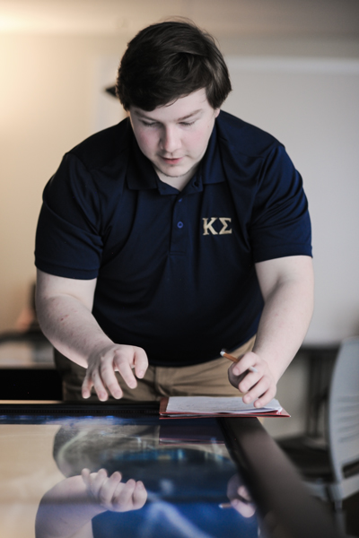
Students are aided by touchscreens on each Anatomage table in the Virtual Anatomy Lab which give the students the ability to zoom in and rotate images with a simple swipe or tap to get a close-up view of bones, muscles, organs, nerves and blood vessels. Furthermore, on Anatomage tables, Life University students can press “reset,” allowing them to explore and learn at their own pace without fear of losing their work.
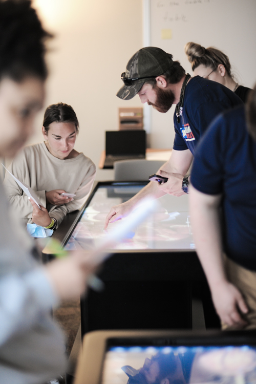
Life University chiropractic students receive all these benefits by using Anatomage tables in the Virtual Anatomy Lab and get hands-on anatomy and physiology education without being exposed to harsh chemicals. This enhances an environment in which students employ problem-based learning and work collaboratively as they learn from each other using the latest technology in our Virtual Anatomy Lab.
From the Virtual Lab
Palpation and Adjustment Trainer (PAT)
RESEARCH & TECHNOLOGY
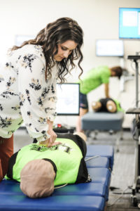
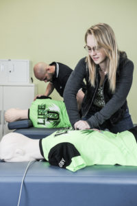
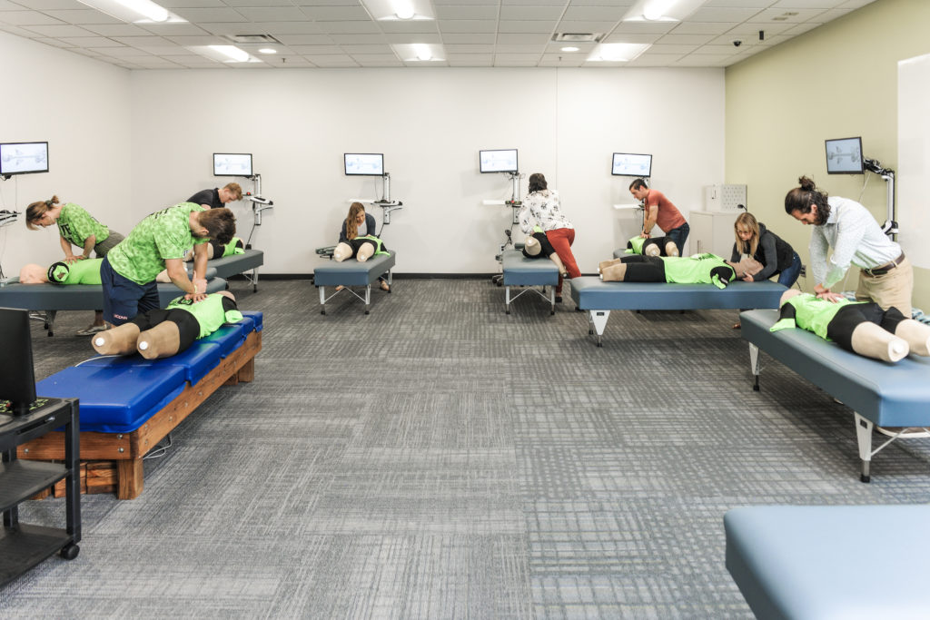
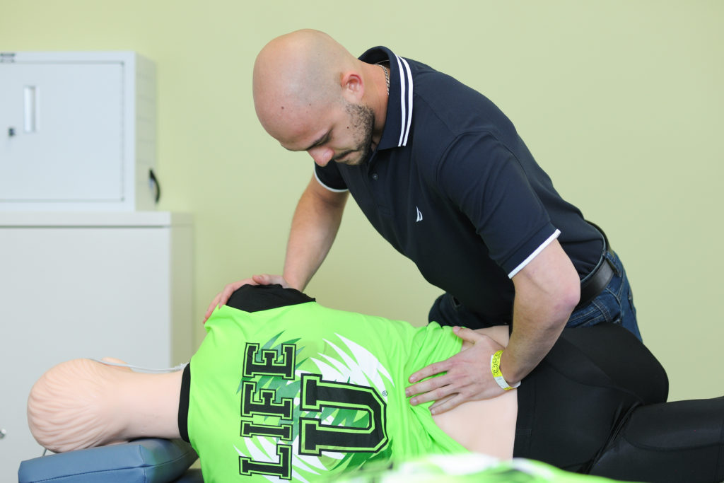
Developed by the Life University Dr. Sid E. Williams Center for Chiropractic Research, PAT (Palpation and Adjustment Trainer) allows Life U Doctor of Chiropractic students to learn the biomechanics of chiropractic adjustments and to develop hands-on skills and techniques prior to beginning the clinical phase of their education.
According to Life University President Dr. Rob Scott, PAT is just the latest technological breakthrough the University has developed as it continues in leading the Vital Health Revolution.
“Imagine for a moment a chiropractic technique lab that brought high touch, high tech innovation to our students, a lab that incorporates technology to help our students through tactile training develop the necessary motor skills to deliver a precise chiropractic adjustment,” Dr. Scott described, adding that PAT is used in addition to – not as a replacement for – adjusting actual patients.
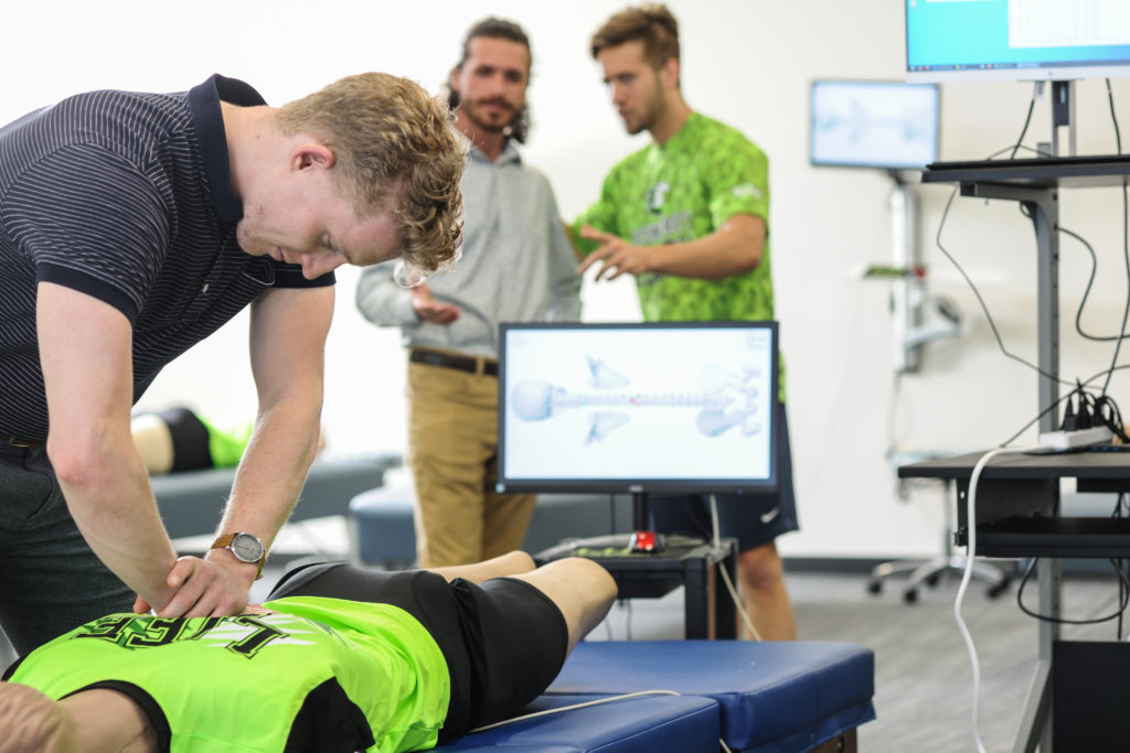
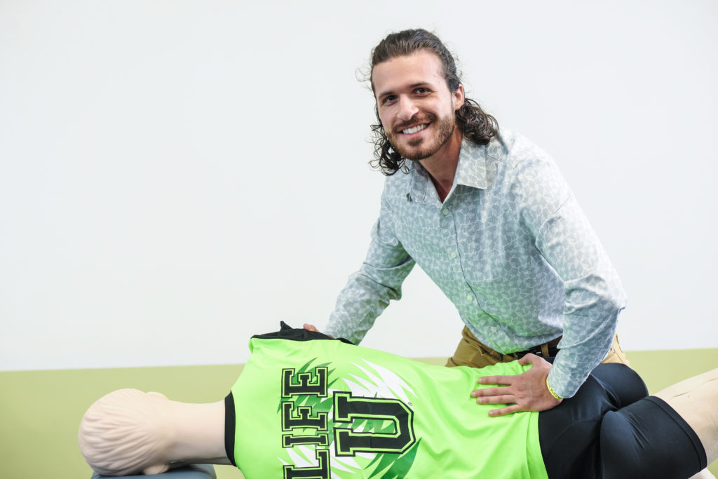
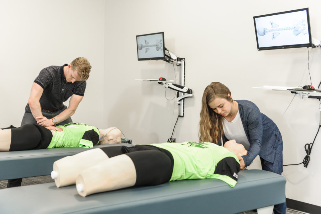
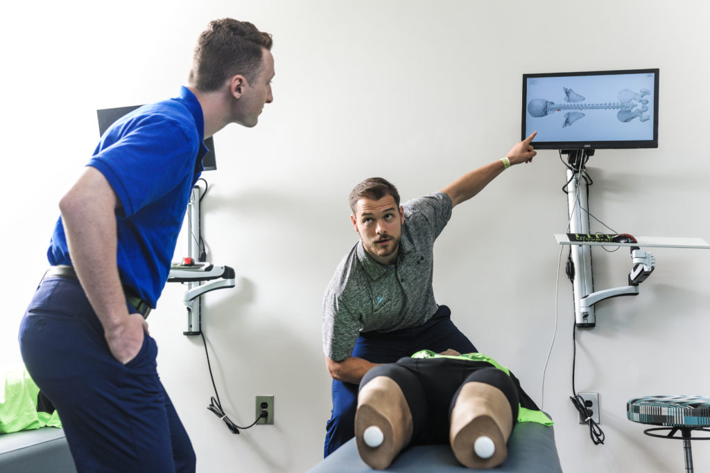
The most advanced palpation and adjusting technology in the Chiropractic profession, PAT is an anatomically accurate, technology-based mannequin with the look, feel, size and weight of an average person. The mannequin features a 3D-printed or molded spine, pelvis and occiput surrounded by viscoelastic skin and soft tissue, simulating the actual experience of adjusting a human patient.
PAT contains 64 embedded pressure sensors at key spinal landmarks from the skull (EOP) to the pelvis and sacrum, helping students learn how to locate vertebral landmarks by palpation, find restricted motion and perform adjustments with controlled amounts of force and speed, along a specific vector.
Along with the force plate technology, PAT gives students immediate, objective feedback about the “where, how hard, how fast and in which direction” part of adjusting, allowing them to develop these important skills necessary for patient care. Computer software monitors pressure levels at each sensor and shows the location of contact, with labels that can be turned on or off.
